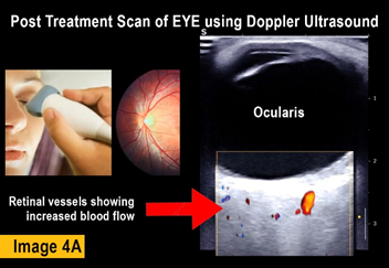
FUNCTIONAL IMAGING CONFIRMS THERAPEUTIC RESPONSE
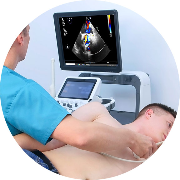
One of the most comprehensive ways to confirm the results of any treatment is by clinically tracking the body's physiological response from underneath the skin. Functional Diagnostic imaging captures measurable data about the injured or inflamed area, allowing both clinicians and patients the ability to identify therapeutic progress in real time. Widely preferred scanning modalities include the Doppler Blood Flow Ultrasound (or sonography) and Elastography, both using high-frequency sound waves to view inside the body. Like an internal video camera, these high speed scanning innovations capture actual movement of the body's internal organs. This offers a vast amount of biometric information about the patient’s condition, in comparison to still images of conventional x-rays. The ultrasound's ability to evaluate abnormalities within the soft tissue in research and clinical trials are widely used in recording evidence-based biomarkers to trace therapeutic efficacy.
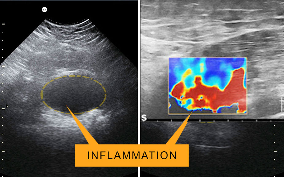 |
DETECTING INFLAMMATION: Part of the complex biological response of body tissues to harmful stimuli (such as pathogens, damaged cells, or irritants) is inflammation. It is also a protective response involving immune cells, blood vessels, and molecular mediators. Detecting the function of inflammation allows us to eliminate the initial cause of cell injury, clear out necrotic cells and tissues damaged from the original insult and the inflammatory process, and initiate tissue repair. The five cardinal signs are heat, pain, redness, swelling, and loss of function. Inflammation is a generic response, and therefore it is considered as a mechanism of innate immunity. Too little inflammation could lead to progressive tissue destruction by the harmful stimulus (e.g. bacteria) and compromise the survival of the organism. In contrast, too much inflammation, in the form of chronic inflammation, is associated with various diseases, such as hay fever, periodontal disease, atherosclerosis, and osteoarthritis.
ACUTE inflammation is the initial response of the body to harmful stimuli from the blood into the injured tissues. A series of biochemical events propagates and matures the inflammatory response within the injured tissue. Prolonged or CHRONIC inflammation, leads to a progressive shift in the type of cells in the inflamed area and is characterized by simultaneous destruction & healing of the tissue from the inflammatory process.
ADDRESS THE STRESS
Analyzing STRESS & ANXIETY from a holistic point of view means identifying the body’s interconnected systems (ie. circulatory, cardiovascular, nervous, lymphatic, endocrine etc.) and its many touch points for stimulation. This analysis should also offer a comprehensive breakdown of the body's HEALING capacity- which includes our hormones, digestive system, immune system, brain, heart-- all the way down to our cells and mitochondria. Stress is part of life, and comes in many forms including physical, emotional, mental and environmental. Foods we eat, unhealthy relationships, difficulties at work, toxins in our environment, even poor posture or lack of sunshine can all create stress on our bodies. But when stress is catastrophic or becomes chronic, it creates imbalances in this functioning that are much more likely to promote disease while at the same time preventing healing from taking place.
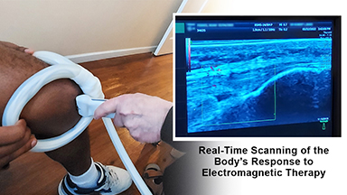
 IDENTIFYING TREATMENT EFFICACY: How much effect is your medicine actually having on the body? This level of clinical reporting provides findings on the immediate physiological effects of any therapeutic solution. Results can be based on a BEFORE‐AND‐AFTER visual response comparison, quantifiable biometrics and a sound description of the clinical imaging ‐ thus aiming to show the body's potential reaction to that device (if any) by virtue of a post‐treatment applied scan. Imaging provides scan studies designed to assess, confirm or challenge (if necessary) any device's claims to confirm the device’s impact on the body. Measurable evidence reports can be conducted through the use of advanced ultrasound scans who seek real-time evidence-based valuation. IDENTIFYING TREATMENT EFFICACY: How much effect is your medicine actually having on the body? This level of clinical reporting provides findings on the immediate physiological effects of any therapeutic solution. Results can be based on a BEFORE‐AND‐AFTER visual response comparison, quantifiable biometrics and a sound description of the clinical imaging ‐ thus aiming to show the body's potential reaction to that device (if any) by virtue of a post‐treatment applied scan. Imaging provides scan studies designed to assess, confirm or challenge (if necessary) any device's claims to confirm the device’s impact on the body. Measurable evidence reports can be conducted through the use of advanced ultrasound scans who seek real-time evidence-based valuation.
|
CANCERSCAN
Specialized Clinical Programs
Diagnostics brings world-class expertise in advanced medical imaging to detect cancer at its earliest, most treatable stages. As a pioneer in high-resolution ultrasound, 3D/4D Doppler imaging, and elastography, Dr. Bard designs targeted scanning programs for prostate, breast, skin, thyroid, and other cancers, tailored to each patient’s risk profile. His protocols integrate cutting-edge technology with evidence-based prevention strategies—identifying tumors, mapping blood flow, and assessing tissue health without radiation exposure.
Beyond diagnostics, Dr. Bard’s imaging programs support prevention and surveillance, enabling early intervention for high-risk patients and monitoring treatment progress with precision. Whether screening for hidden cancers, evaluating suspicious lesions, or conducting follow-up scans, Bard Diagnostics delivers clarity, accuracy, and peace of mind. Patients and physicians worldwide trust Dr. Bard for his ability to transform complex imaging data into actionable, life-saving insights—helping stop cancer before it advances. |
 |
 |
 |
 |
 |
 |
 |
 |
 |
|
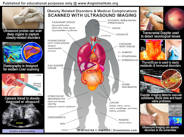
Imaging to Detect Obesity-Linked Health Disorders
Obesity is a long-term health condition impacting more than a billion people worldwide, with numbers rising steadily due to modern dietary patterns, low physical activity, genetics, and environmental influences. Beyond visible weight gain, obesity disrupts nearly every organ system, triggering widespread complications that shorten lifespan and degrade quality of life. These include cardiovascular problems, type 2 diabetes, liver damage, hormonal and neurological disturbances, certain cancers, and musculoskeletal disorders. While traditional diagnostic approaches often involve invasive procedures or costly imaging scans, ultrasound technology has become an essential, safe, and highly adaptable method for evaluating obesity-related diseases. The poster provided illustrates how ultrasound can scan multiple organ systems affected by excess weight, supporting early detection, risk evaluation, and targeted treatment planning.
HOW OBESITY IMPACTS ORGAN SYSTEMS: Obesity is not confined to fat accumulation beneath the skin. It causes hormonal imbalance, inflammation, and structural strain that interfere with normal organ function. Key systems affected include:
1. Cardiovascular Health
2. Liver Function
3. Pancreatic Health
4. Gallbladder Disorders
5. Brain and Neurological Function
|
6. Thyroid and Hormonal Balance
7. Reproductive Health
8. Cancers Associated with Obesity
9. Musculoskeletal Stress
10. Circulatory and Skin Conditions |
(See complete studies and reports on Obesity Imaging)
|
|
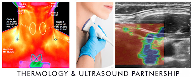
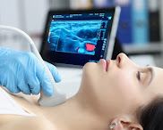
THYROIDSCAN
For decades, the diagnostic gold standard for parathyroid adenomas has been technetium-99m sestamibi scintigraphy—a nuclear medicine technique boasting a reported accuracy of approximately 95% in experienced centers. However, its dependence on radioactive isotopes and its limited capacity for precise anatomical localization have motivated researchers to seek alternatives. Dr. Robert L. Bard, a leader in advanced ultrasound imaging, has introduced a breakthrough protocol that leverages high-frequency ultrasound and 3D Doppler technologies for non-invasive parathyroid evaluation. His methodology offers not only diagnostic accuracy but also a path to image-guided interventions that may transform the treatment landscape.
Endocrinologists embrace cutting-edge imaging to transform diagnostics and patient outcomes:
For millions struggling with thyroid disorders, early detection and accurate monitoring can make all the difference. From benign nodules to aggressive cancers, subtle changes within the small butterfly-shaped gland in the neck can have systemic effects. In this landscape, ultrasound imaging is emerging as the gold standard—redefining how endocrinologists assess, diagnose, and guide treatment. .(see complete tech details)
For patients, the shift to in-office ultrasound offers significant benefits:
✅ Instant results reduce anxiety and expedite care plans.
✅ Dynamic imaging allows them to watch and understand findings in real time.
✅ Fewer referrals and delays, which means more efficient care.
For complete details, visit THYROIDSCAN.org
|
|
MENOSCAN
Menopause Care & Precision Diagnostics
MenoScan.org represents a groundbreaking initiative led by Dr. Robert Bard, bringing together top medical specialists to redefine the way menopause is understood, diagnosed, and managed. Menopause is far more than a shift in reproductive function—it is a complex, multi-system transition that impacts hormonal balance, bone density, cardiovascular health, neurological function, and overall metabolic stability. Traditional approaches often address menopause symptomatically, but MenoScan offers a deeper, science-driven strategy: leveraging advanced medical imaging to uncover the hidden physiological changes that occur during this pivotal stage of life.
 |
Through strategic partnerships, Dr. Bard collaborates with leading endocrinologists to assess and monitor hormone levels and thyroid function, providing a crucial window into the endocrine shifts that drive menopausal symptoms. Orthopedic specialists join the program to investigate bone loss and muscular changes, helping women mitigate risks such as osteoporosis and age-related frailty. Cardiologists contribute expertise in detecting early vascular and heart conditions, recognizing that menopause significantly raises cardiovascular risk factors. These multidisciplinary collaborations ensure that MenoScan covers the full spectrum of health concerns women face in midlife and beyond.
What sets MenoScan apart is its commitment to advancing therapeutic options through precise, non-invasive imaging. By identifying changes in tissues, glands, blood flow, and structural integrity long before symptoms escalate, MenoScan empowers both patients and physicians to make informed, personalized decisions about prevention and care. This program bridges the gap between conventional menopause management and the next generation of women’s health innovation, ensuring that no aspect of this life stage goes unexamined. MenoScan is not just a diagnostic tool—it is a movement toward proactive, integrative menopause care. Through its collaborative network of medical experts, it is setting new standards for understanding and addressing the multifaceted effects of menopause on the female body. Our program offers diagnostic imaging focused on:
| CARDIOVASCULAR DISEASE (CVD) |
BREAST AND REPRODUCTIVE HEALTH |
CLINICAL IMAGING OF MENOPAUSE-RELATED HAIR LOSS |
| OSTEOPOROSIS AND BONE HEALTH |
COGNITIVE DECLINE AND BRAIN HEALTH |
HORMONAL & THYROID FUNCTION |
| METABOLIC SYNDROME AND WEIGHT GAIN |
|
|
Visit: MENOSCAN.ORG for complete details and an expanded study on Midlife Health programs
|
CLINICAL RESEARCH PROJECTS
The concept behind PERFORMANCE TESTING underscores the core commitment of any health-related product to “ensure that health or medical devices shall consistently and safely provide the effects and benefits they are intended. Dr. Robert Bard (NYC) officially drafted an official update to his blueprint coordinating clinical monitoring and validation of non-invasive devices through diagnostic imaging protocols (under Institutional Review Board approved through HHS and FDA regulations). The primary monitoring and biometric reporting research protocol employs the use of medical grade 3D Doppler Ultrasound imaging, Elastography and other non-radiation based imaging solutions. (PDF:see details)
 |
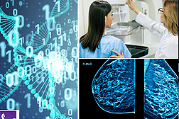
RESEARCH PROJECT: DENSE BREAST IN ATHLETIC COMMUNITY-
9/22/2021- For women with any level of breast density, one of the major concerns is the alarming rate of false positives that may align with cancers missed by a mammogram. The other concern is that women with dense breasts have a naturally higher risk of breast cancer than women with fatty breasts, and the risk increases with increasing breast density. (This increased risk is separate from the effect of dense breasts on the ability to read a mammogram.). The main focal points of this research project covers the diagnostic study of ATHLETIC WOMEN or those with LOW BODY MASS INDEX (bet 12-22% body fat).
ACTIVE SURVEILLANCE
Where Recurrence Prevention Scans are implemented after cancer treatment or surgery, Active Surveillance is often recommended once you are diagnosed of an early stage cancer- one that is identified as slow-growing such as prostate, breast, kidney, and thyroid cancer. Monitoring cancers during its very early stages (instead of immediate treatment) may be a good option. This means closely watching a patient’s condition but not giving any treatment unless there are changes in test results that show the condition is getting worse. Active surveillance may be used to avoid or delay the need for treatments such as radiation therapy or surgery, which can cause side effects or other problems. During active surveillance, certain exams and tests, such as blood tests, imaging tests, and biopsies, are done on a regular schedule to monitor the condition. Active surveillance may be used in certain types of prostate cancer and in some other types of cancer. It is a type of expectant management. (See complete report)
Contributing to the evolution of SCIENCE
by: Dr. Robert L. Bard
 |
I welcome all solutionists and innovators who support the healing arts community. I have devoted my specialized work to pursue the advancement of MEDICAL RESEARCH with NON-INVASIVE SURROGATE ENDPOINTS with the hopes of contributing my talents to the performance of your current and future projects. With over 45 years in the field of advanced diagnostic science, my life's work has been about the clinical examination and targeted analyses of all subdermal disorders using the latest quantifiable digital imaging innovations. I have established an entire foundation dedicated to Medical Research committed originally to the exploratory studies of all cancer treatment proctocols. Alongside this, I have also been most active in collaboration with some of the top treatment strategists, health centers, clinical labs, experimental / alternative treatment professionals and medical device manufacturers. I have earned a reputation for my investigative approach within various examination paradigms including progress monitoring and surveillance.
As a CO-INVESTIGATOR, I have the greatest interest in supporting all entities committed to contributing technical innovations and new advancements in treatment solutions for our medical community. By this, I wish to build a partnership with establishing research teams to conduct clinical test projects where my talents to support visual reporting through imaging to be a priceless benefit to your overall objectives. Most research sponsors and medical developers' testing and tracking needs often fall into one of a number of common task categories- all within the objectives of public health, safety and healthcare support. I look forward to learning about your project with the hopes of supporting your needs and objectives.
|
THE NON-INVASIVE MOVEMENT:
AVOID THE RISKS OF CUTTING & UNNECESSARY BIOPSIES
When it comes to finding abnormalities in a patient exam, many conventional-minded doctors tend to tread on the side of caution... but usually at YOUR expense! They find an unusual spot that appears questionable and their first reaction is to cut it out and send it to the lab for a BIOPSY. As with all invasive surgical procedures (however large or small) carries risks including bleeding, infections, post-surgical scars and potential damage to nearby tissues and organs.
The year is 2019- the era of non-invasive technologies where identifying what's under the skin no longer needs to be about cutting into it. The age of robotics, artificial intelligence (AI), highly developed laser applications and advanced sonic diagnostic protocols are all fast replacing the age-old scalpel as part of risk reduction, time/cost advantages and increased performance in the world of clinical diagnostics and medical treatment.
Imaging technologies like the 4D Doppler Ultrasound™ can accurately and successfully scan, study and fully diagnose any skin anomalies and sharply view what's going on underneath. Derm imaging specialists stand on the side of innovation as they confidently rely on the most current devices to deliver the most accurate readings while bringing significant reduction to patient stress under a scan- that takes mere minutes!
"Before & After" Studies
The most sensible and logical way to identify the results of any treatment is by tracking the body's response to it. Controlled testing must show the patient's condition PRE and POST effects, where true data-finding is collecting the necessary EVIDENCE of its claims. The investigator can pull a significant amount of data from this form of oberservational testing and recording: including stage-by-stage bodily response to future projections of possible side effects. Recording of any and all psysiological response means the researchers are counting on the patient's body to tell us what it is undergoing during the testing phase. To prevent mis-reading and erroneous reports, trials tend to work with a large number of test patients (commonly 50-100) and may also employ redundancies like undergoing multiple testing protocols for a second or even third opinion. To capture the benefits of a BEFORE AND AFTER review, Imaging is often used as a standard screening solution for the response of most of the major organs.
|
"SEEING IS BELIEVING":
Advantages of Imaging in Research Studies
 QUANTIFIABLE DETECTION OF THERAPY RESPONSE QUANTIFIABLE DETECTION OF THERAPY RESPONSE
MEDICAL IMAGING refers to the set of FDA approved modalities that are used to view the human body in order to diagnose and monitor medical conditions or the response of its treatments. The types of common modalities include X-rays, MRI (magnetic resonance imaging), Ultrasound, CT scan (computed tomography scan) and Nuclear medicine imaging including Positron Emission Tomography (PET) [1][2]
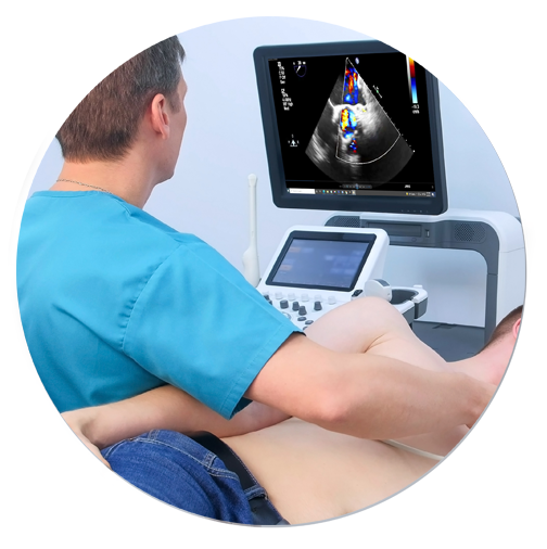
Ultrasound imaging (sonography) uses high-frequency sound waves to view inside the body. Because ultrasound images are captured in real-time, they can also show movement of the body's internal organs as well as blood flowing through the blood vessels. Unlike X-ray imaging, Ultrasound uses NO ionizing radiation exposure whatsoever.[3] This modality (sometimes referred to as "the orthopedic stethoscope"), offers easy access, portability, affordability and real-time monitoring and is safe for all patients, including those with cardiac pacemakers and metal implants, without any contraindications.
TRACKING THERAPEUTIC EFFECTS THROUGH BLOOD FLOW REACTIONS
To date, the ultrasound's ability to evaluate abnormalities within the soft tissue such as cysts, tumors and inflammation is used to help identify an expanded set of pathologies in the body. Since the early 1970's, Dr. Robert L. Bard (NYC Cancer Radiologist) has used diagnostic imaging for pre and post procedural guidance, and diagnostic care (screening and monitoring) of his patients. Dr. Bard is also recognized for his use of ultrasound in pharmaceutical research and clinical trials, where his leadership in analytical interpretation is sought after worldwide for identifying markers and therapeutic efficacy.
Throughout his career, Dr. Bard has employed this imaging strategy to detect, track and confirm the body's reaction to a variety of therapeutic interventions. He has conducted medical center based double-blinded, corporate sponsored and private studies reviewing the effects of injectable therapies (PRP, Stem Cell therapies, etc) as well as non-invasive therapeutic interventions in studies of neuro-stimulation, electrostimulation and electromagnetic field treatments. His approach involves the comparative study of measurable scanning data or quantitative ultrasound (QUS) which aims at recording interactions between the behavior and activity of biological tissue microstructure and ultrasound waves [5][6]. From a time-based comparative study of the treated area (before and after studies), Dr. Bard applies the use of blood flow detection technology or hemodynamic data gathering protocols, document specific objective and quantifiable biological responses to therapeutic treatments.
Ref:
1) Medical Imaging/FDA: https://www.fda.gov/radiation-emitting-products/radiation-emitting-products-and-procedures/medical-imaging#:~:text=Medical%20imaging%20refers%20to%20several,monitor%2C%20or%20treat%20medical%20conditions.
2) https://sonosimaging.com/press-corner/understanding-the-different-types-of-imaging/
3) https://www.fda.gov/radiation-emitting-products/medical-imaging/ultrasound-imaging
4) Diagnostic imaging to detect and evaluate response to therapy in bone metastases from prostate cancer: current modalities and new horizons https://pubmed.ncbi.nlm.nih.gov/26956538/ |
|
IRB-BASED VALIDATION STUDY / VISUAL AND QUANTIFIABLE REPORTING OF NON-INVASIVE THERAPEUTICS THROUGH MEDICAL IMAGING PROTOCOLS
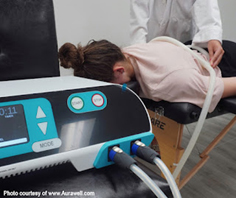
By the year 2000, the vast majority of non‐invasive wellness devices on the public market have deep rooted themselves into the WELLNESS and FUNCTIONAL communities, leading to the sales of ‘invisible treatments’. These devices employ one of a number of technology based modalities including: pulsed electromagnetic frequency, biofeedback, shockwave sonic pulse as well as light‐related energy therapies like cold laser, blue light and infrared just to name a few.
The concept behind ‘MEDICAL VALIDATION’ underscores the complex commitment of “ensuring that the medical device being manufactured will consistently provide the intended benefits for its use condition. Clinical validation is usually done through a series of tests and inspections.” [1] To conduct this medical validation officially can be offered through various protocols, an Institutional Review Board approved by the HHS and FDA regulations. One protocol that can be available for consideration by the IRB panel is the use of medical grade 3D Doppler Ultrasound imaging. The use of medical imaging technologies like an ultrasound alongside the interpretation of a certified clinical radiologist may offer biometric assessment and analyses of all scanned readings and the collection of ample data to confirm or validate a health device’s effects on the body as marketed.
 |
PERFORMANCE TESTING & VALIDATION PROGRAM MAY CHALLENGE OR SUPPORT PUBLIC CLAIMS
Especially when a product is promoted as a health‐related solution, testing is an important and highly critical step from a legal standpoint. This step is directly involved in a developer’s statements and claims about the device. Before any device is to be promoted for a specific pathology, a series of formal performance exams must be conducted to suit regulatory acceptance by an outside agency or clinical team approved by a federally backed review board. Considerations for validation include: range of physiological effects, warnings about the possible hazards, physical risk to the user in regular use, adverse reactions (potential side effects)‐ and all these elements are from data acquired during a validation study [2][3]. To confirm (or validate) that a product is effective within the statements and claims that it provides the public, the manufacturer would need to invest in an exclusive and dedicated lab or agency that is poised to perform this level of testing. Said lab must acquire enough data (according to the IRB board) to supports the aspired claims. If/when enough data has proven the hypothesis true, this would add to the product’s marketing credence as well as confirms the device’s ability as a health or wellness product. Any other type of observational testing outside of the authority of an IRB is considered ANECDOTAL and must be established as such‐ which holds no credence as an industrywide claim whatsoever. |
|
Recent & Innovative PROJECTS
Within the past decade, the world of medicine has witnessed (and achieved) a significant amount of success with STEM CELL therapies managing a wide range of disorders. There are 2 main types of stem cell transplant processes- AUTOLOGOUS (source of harvested cells are the same as the recipient) and ALLOGENIC (sourced from a matching donor). Concentrates of regenerative cells can be extracted from bone marrow, unbillical cord (newborns) or fat (adipose) cells & bloodstream. Patients are (now) recognized to receive treatments for a growing list of isses such as ORTHOPEDIC damage (bone and joint injuries) and AUTO-IMMUME DISORDERS (lupus, arthritis, diabetes ). NEUROLOGICAL ISSUES are also now being addressed by stem cell treatments whereby research and testing for treatment efficacy relating to the Nervous System is also in full swing within the clinical community.
Dr. Bard's 4D Doppler
capabilities have become the preferred non-invasive solution with research programs to seek out treatment testing or application performance tracking for this exact type of exploratory testing. Dr. Bard has the capacity to identify and track prestroke vasospasms, any increase in intracranial pressure, brain aneurysms
and many degenerative disorders. (See related article) |
Early collaboration with technological FRONTIERSHIP
 |
In 1976, during Dr. Bard's days as a young radiologist, he was approached by Dr. Henry Leis Jr., the pioneer doctor who wrote the very first text on breast cancer and developed mammography 18- a means of early diagnosis and instrumental in the use of many of the less invasive procedures used in the treatment of breast cancer today. He confessed with great concern that he had all these patients with lumpy or cystic breasts developing tumors that he could clearly feel but the mammogram kept missing it. Seeking Dr. Bard's help through sonogram technology, they worked on his patients together and the sonogram clearly identified and quickly diagnosed a mass as either a cancer or a benign cyst, in a dense, lumpy breast. Since then, the sonogram became incorporated in high-risk patients’ regimen every six months religiously because it finds tumors while they're small and “lumpectomy” surgery is curative if the mass is less than 1 cm. This is alongside doing mammograms once a year in women over 50 or unless they have a history of cancer- at which case, we do it starting at age 45. |
|
|